Ready for Lift Off: 3D Scanning in Anatomical Pathology
Discover the future of pathology with advanced 3D models, innovative teaching methods, and improved patient care through cutting-edge technology.
Impact Innovation | Innovarium

In the pursuit of health care innovation, the Innovarium Launchpad offered a vital opportunity for creative thinkers at Providence Health Care (PHC). The initiative provides seed funding to support groundbreaking ideas with the potential to transform the health care system.
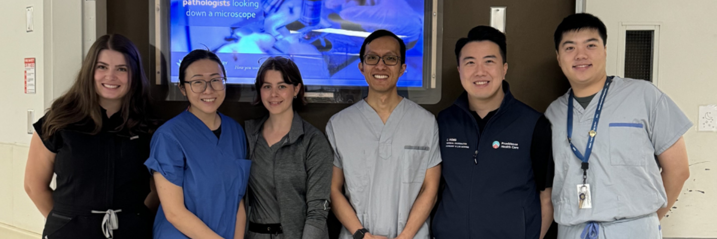
Among the three projects supported by the Launchpad is 3D Scanning in Anatomical Pathology, led by Kaelyn Wagland, a Pathologists’ Assistant, alongside her dedicated team, including Helena Froberg, Crystal Leung, Richard Li, Chris Wong, Bobby Grewal and Dr. Lik Lee. Their innovative approach addresses a fundamental limitation in the field: Human anatomy and histopathology are inherently the studies of 3D objects. Learning the relationships between these 3D structures is of utmost importance and yet, pathology’s diagnostic and education tools remain to be represented in a 2D plane.
Transforming Anatomical Pathology
“Our innovation focuses on integrating 3D surface scanning technology into anatomical pathology labs,” explains Wagland. “Traditional 2D digital photography captures limited angles and depths of pathology specimens. With 3D scanning, pathologists can capture highly detailed, fully manipulatable 3D models of specimens, providing a clearer, more comprehensive view. This innovation will enhance clinical accuracy and improve teaching methods.”
The need for this advancement is clear: examination of surgical specimens is critical to patient care, yet traditional photographic methods don’t quite capture the full spectrum. 3D scanning offers a revolutionary leap in how pathology specimens are captured and shared. With 3D scanning, pathologists can capture highly detailed, fully manipulatable 3D models of specimens.
The benefits of 3D scanning extend beyond solely improved diagnostics. The technology also promises greater learning opportunities for residents and PA students, as well as cost savings. Digital models reduce the need for physical specimen storage, potentially lowering the overall cost of handling. Moreover, the portability of these digital models facilitates easier sharing across health systems, promoting interprofessional collaboration.
The Benefits Beyond
Currently in the proof-of-concept stage, this innovative 3D scanning project is being piloted at the Foothills Hospital Anatomical Pathology Lab in Calgary. The collaboration with Foothills Hospital is key to refining the technology, as their team has provided valuable insights into scanner setup and implementation. While the technology itself is not new—3D scanning has been used extensively in fields like engineering—it is still in its early stages in anatomical pathology. In fact, the use of 3D models in pathology is so novel that it has the potential to change the field by offering a more detailed and manipulable way to capture and study pathology specimens compared to traditional 2D photography.
The team at PHC is building on this proof of concept and exploring how 3D scanning can improve not just diagnostic accuracy, but also the overall experience of pathologists. Looking ahead, the project team envisions a future where 3D scanning is integrated into pathology labs nationwide. “Receiving funding for a clinical 3D scanning project gives us the opportunity to advance healthcare technology and improve patient outcomes through innovative solutions,” Kaelyn Wagner reflects. “It empowers the development of accurate diagnostic tools and fosters collaboration, ultimately translating visionary ideas into impactful health care practices.”
The technology has potential applications across a variety of fields, from medical education—where it can help students and residents better understand complex anatomical structures—to patient care, where it can be used to create 3D models that improve communication between patients and doctors. Imagine, for instance, a patient holding a 3D-printed model of their heart after a transplant, helping them better understand their surgery and recovery. Additionally, 3D scanning could enhance collaborative efforts across health care systems, allowing for easier sharing of complex cases during specialist rounds or medical conferences.
This project has the potential to change the way specimens are handled, documented, and studied. By marrying cutting-edge technology with clinical expertise, the integration of 3D scanning could set a new standard in specimen imaging, enhancing diagnostic accuracy, educational outcomes, and research capabilities.
Stay Tuned for More Launchpad Innovations
Looking ahead, Innovarium is committed to driving programs forward that will continue to expand and flourish at the new St. Paul's and the Clinical Support & Research Centre. Projects like these will be supported by state-of-the-art facilities and programming, fostering an environment where innovation and collaboration can thrive.
The journey of innovation at PHC doesn’t stop here. We invite you to return soon to learn about our other two funding recipients from the Innovarium Launchpad coming soon to Connect.